
Bone mineral density of spine and hip was measured by DXA
The accuracy of DXA in measuring different anatomical parts of the human body varies [4-7]. The accuracy of DXA in measuring the spine is 0.5%~2%, but usually >1%. The accuracy of the hip is 1% ~ 5%, with the femoral neck and big rotor (1% ~ 2%) is superior to the Ward ‘s triangle (2.5% ~ 5%) (4. 6. 8). Despite the high content of cancellous bone in Ward’s triangle and its high sensitivity to changes in BMD [9], its poor accuracy due to its small projection area and sampling and repeatability errors limits its clinical application. To minimize the impact of scanning position on accuracy when performing DXA measurements, the hips and knees were flexed on a support to allow the spine to be true on the platform to reduce lumbar lordosis during the determination of the BMD in the anteroposteric lumbar position (posteroposteric PA). During the hip scan, the thigh was slightly abduction and pronated, and with the help of a postural fixation device, the femoral neck was positioned parallel to the scanning table to avoid increasing BMD due to shortening of the femoral neck (reduced volume for the same bone mineral content). In the determination of hip BMD by DXA, different leg positions can cause significant errors, ranging from 0.9% to 4.5% for femoral neck, 1.0% to 6.7% for Ward’s triangle, and 0.4% to 3.1% for greater trochanter [6]. Therefore, when DXA scans the hip, correct posture will significantly reduce the error, which is the key to ensure good precision Angle.
If the results of hip BMD measured by DXA are not consistent with clinical manifestations, one should be performed
The author should check whether the scanning position is correct; On the other hand, clinicians should consider the influence of scanning position on BMD. In addition to the influence of position on the accuracy of DXA measurement, other reasons may also affect the measurement results. Spinal alignment was determined by DXA.
Spinal BMD is defined as the density of the entire vertebral body area, including the vertebral body and arch (cortical bone to cancellous bone ratio 50:50), aortic calcification, degenerative osteoarthrosis, osteopanthogenic spinous process, callus, and compression fractures, which all contribute to increased bone mineral density. However, degenerative changes such as hyperosteoplasia are very common in the elderly over 70 years of age, with a prevalence of more than 60%, which limits the practicality and sensitivity of DXA spine orthotopic measurement in the elderly population. The incidence of osteoporosis is high and serious in middle-aged and elderly people
It is a common disease of old age that threatens the health of middle-aged and elderly people. In order to eliminate the influence of the above factors, the development of DXA lumbar lateral scanning technology (1121, the early DXA scanner for other lumbar scanning, the disease is prone to maintain the position of the scanning, which is
Affected the accuracy, which was 2.8% to 5.9%!
At the same time for some diseases
People, especially those with severe osteoporosis, have difficulty turning over.
In recent years, DXA scanner adopts fan-shaped beam rotating “C” shaped arm scanning, allowing disease
The spine BMD was measured anteroposterically in supine position and the C-arm scanner was rotated 90°
The patient can be measured by DXA in the lateral position of the reputation column without moving
The accuracy of lateral measurement was 1.6% in normal subjects and 2% in patients with osteoporosis. The ideal lateral DXA measurement should analyze the BMD of 4 lumbar vertebrae (L1-L). However, L1 and L4 may be covered by ribs and L4 is obviously overlapped by the pelvic bone. For some patients, only L3 BMD can be analyzed. The ROIS(region of interest) can also be located in the center of the vertebral body rich in cancellous bone (cortical bone/cancellous bone ratio of 10:90), making DXA measurements more sensitive to changes in BMD in lateral than in frontal view. Lateral DXA is used in healthy subjects with columnar osteoporosis (vertebral compression fractures)
The discrimination between bone mass loss caused by corticosteroids is better than that of PA-DXA, which improves the ability to distinguish vertebral fractures from non-fractures [15]. Although DXA has made great progress in measuring spinal BMD. However, for scoliosis, severe humpback and abnormal spinal segmenting [4,61], the operation of DXA scanning is difficult, affecting the accuracy of DXA determination and limiting the clinical application of DXA. Further studies are needed to compare the “volumetric” BMD (mg/cm3) calculated by combined frontal and lateral DXA measurements with the QCT method.
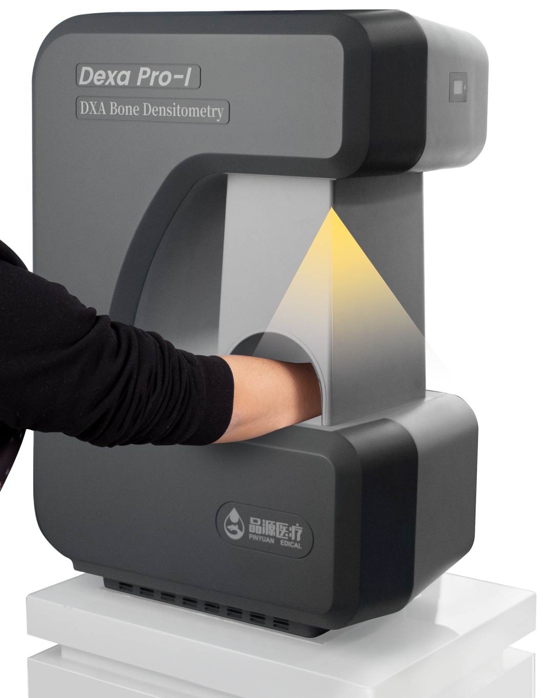
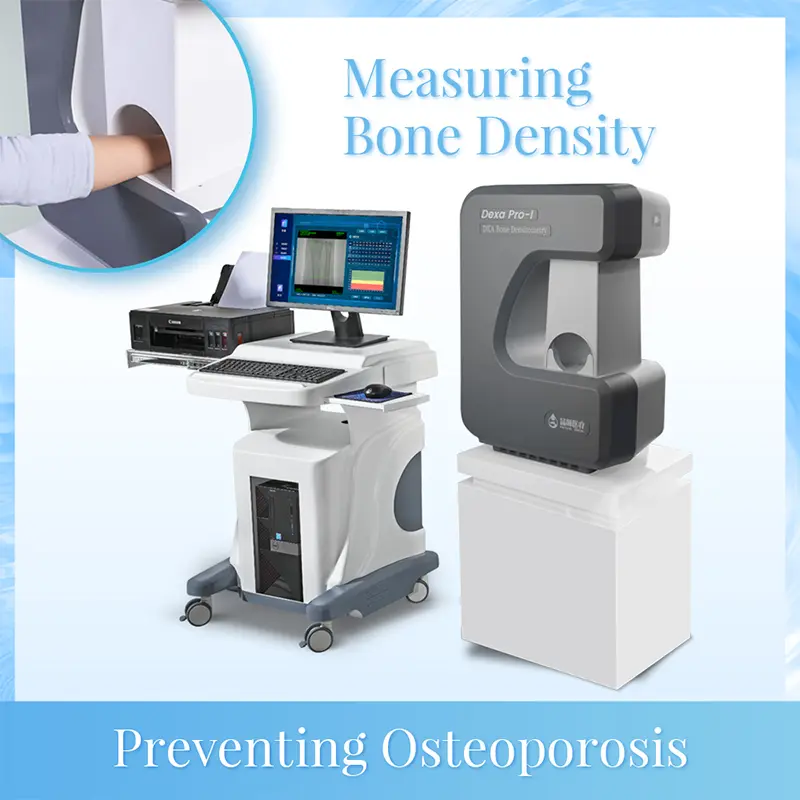
Determination of forearm BMD and body composition by DXA
DXA is increasingly used to determine forearm BM[17]. BMD measurements were performed in the distal radius (cancellous predominance), middle and middle, and distal third of the radius (cortical predominance) with the patient sitting on a chair adjacent to the scanning platform with the forearm positioned on the platform and the hand fixed on the platform with anterior rotation. Whole body bone densitometry can also be performed. This provides a systematic comparison of whole-body BMD and local BMD. To analyze and explore the relationship between systemic BMD and local BMD, and find out the sensitive site of bone densitometry, so as to provide the best choice for clinicians. The accuracy of whole body BMD measurement is 3% to 8%. 19] The accuracy of forearm BMD is 0.8%-13%. Because the accuracy of DXA whole-body BMD is less than that of other parts, the bone is thin
Loose is generally not the preferred scan site for diagnosis. The results of whole body scanning were analyzed by software information system of appropriate human tissues (lean muscle and fat mass), and the results of body composition determination were obtained by DXA. The correlation between the results of body composition determination and other indirect weight measurement methods was good. It is an important field that can be further studied.
Post time: Aug-10-2022