
Osteoporosis is a common bone disease, especially prevalent among middle-aged and elderly people and postmenopausal women. Early detection of bone mass loss is crucial for preventing fractures, and ultrasound bone density examination is precisely a safe and convenient screening method. So, which parts of the body are usually tested by ultrasound bone density examination? What are its principles and advantages? Today, let’s take a detailed look.
Which parts of the body are tested by ultrasound bone density examination?
Ultrasonic bone densitometers usually assess bone density by measuring the sound wave velocity (SOS) and broadband ultrasonic attenuation (BUA) of bones. Common detection sites include:
The radius and ulnar (forearm) : The distal end of the radius near the wrist is a commonly used detection point, which is convenient to operate and suitable for initial screening.

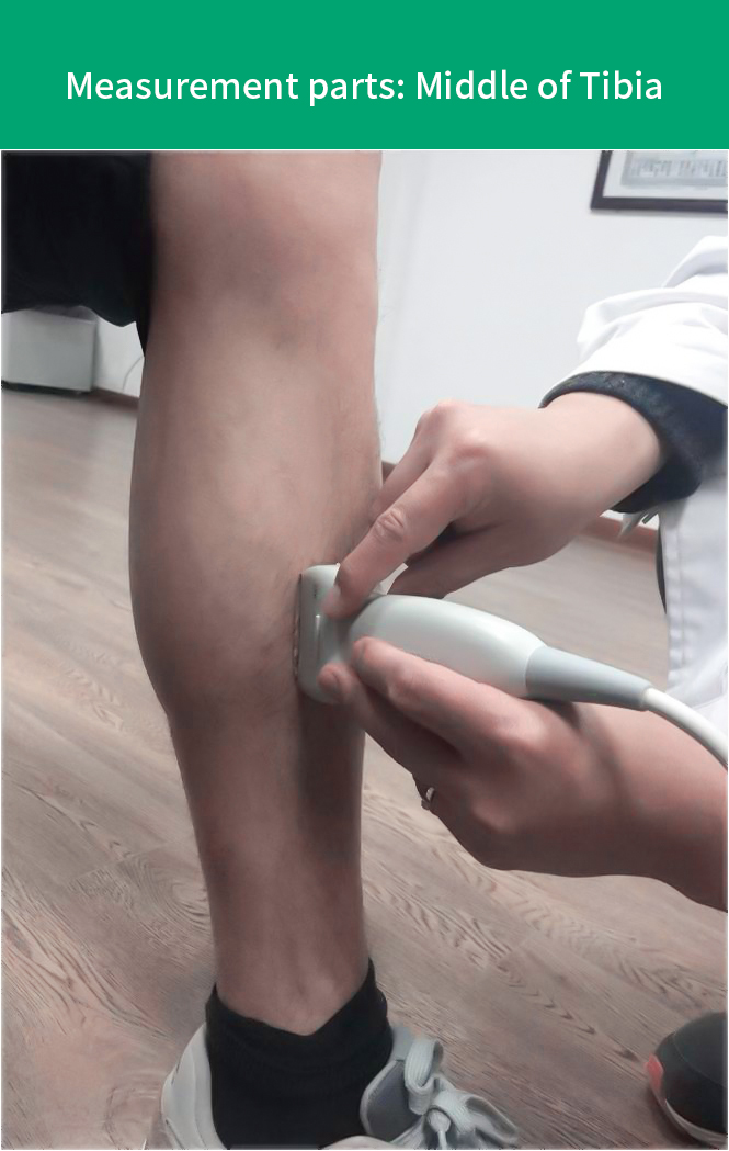
Tibia (lower leg) : The area of the tibia near the ankle joint can also be used for measurement and is suitable for certain special groups of people (such as those who are obese or have lumbar spine disorders).
2. The principle of ultrasound bone density examination
The ultrasonic bone density meter calculates the hardness and density of the bone by emitting high-frequency sound waves through the bone and measuring the propagation speed and attenuation degree of the sound waves.
Sound wave speed (SOS) : It reflects the elasticity of bones. The faster the speed, the higher the bone density.
Broadband ultrasound attenuation (BUA) : It reflects the microstructure of bones. The smaller the attenuation, the better the bone quality.
Combining these two parameters, the risk of osteoporosis can be evaluated. However, it should be noted that ultrasound examination is generally used for initial screening rather than final diagnosis.
3. Advantages of ultrasound bone density examination
Compared with the traditional DXA test, ultrasound bone mineral density examination has the following advantages:
✅ Radiation-free: Suitable for pregnant women, children and patients requiring frequent monitoring.
✅ Easy to operate: Fast detection, usually only a few minutes, no special preparation required.
✅ Strong portability: Some devices can be moved (e.g. Pinyuan BMD-A3), suitable for screening and physical examination.
4. Who needs to have an ultrasound bone density test?
The following groups of people are recommended to have regular bone density screening:
Postmenopausal women (The decline of estrogen accelerates bone loss)
Middle-aged and elderly people over 50 years old (age is the main risk factor for osteoporosis
People who have been taking hormone drugs for a long time (such as glucocorticoids)
Those with a history of fractures or a family history of osteoporosis
Those who lack exercise for a long time or are malnourished
5. How should the inspection results be interpreted?
The results of ultrasound bone mineral density are usually expressed as T values or Z values:
T value: Compared with the bone density of healthy young people, T≤-2.5 indicates osteoporosis.
Z value: Compared with peers, a lower Z value may indicate secondary osteoporosis (such as hyperthyroidism, kidney disease, etc.).
If the screening results are abnormal, the doctor may recommend further DXA tests or blood tests (such as calcium and vitamin D levels).
6. How to prevent osteoporosis?
Even if the bone density test results are normal, bone loss should still be actively prevented.
Calcium and vitamin D supplementation: Milk, soy products, deep-sea fish, etc. are rich in calcium. Appropriate sun exposure can promote the synthesis of vitamin D.
Regular exercise: Weight-bearing exercises (such as walking and rope skipping) can stimulate bone growth.
Avoid bad habits: quit smoking, limit alcohol intake, and reduce the consumption of carbonated beverages.

Ultrasound bone density examination is a safe and rapid screening method for osteoporosis, especially suitable for health check-ups and early risk assessment. If the test results are abnormal, one should seek medical attention promptly, undergo further examinations such as DXA, and take intervention measures to reduce the risk of fractures.
Post time: May-14-2025